39 chest x ray with labels
VinDr-CXR: An open dataset of chest X-rays with radiologist annotations ... Each scan in the training set was independently labeled by 3 radiologists, while each scan in the test set was labeled by the consensus of 5 radiologists. All images are in DICOM format and the labels from training and test sets are made publicly available. Background CheXpert Dataset | Papers With Code The CheXpert dataset contains 224,316 chest radiographs of 65,240 patients with both frontal and lateral views available. The task is to do automated chest x-ray interpretation, featuring uncertainty labels and radiologist-labeled reference standard evaluation sets. Source: Deep Mining External Imperfect Data for Chest X-ray Disease Screening
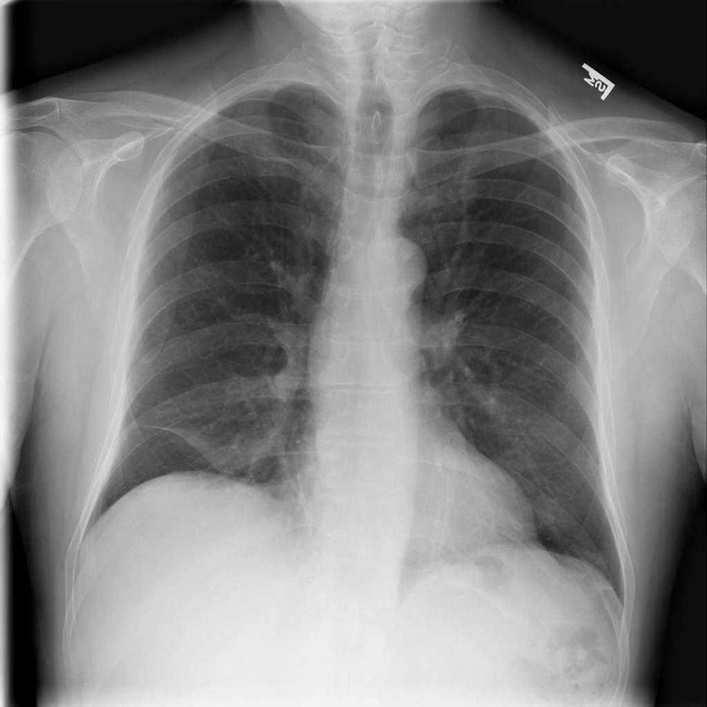
Chest X-Ray's Role in Asthma Screening and Diagnosis A chest X-ray points the X-ray beams towards the chest to take a picture of your lungs and chest area. A chest X-Ray shows: Lungs Heart Several major blood vessels in the chest Ribs (bone and metal, which are dense, show up white on X-rays) The air in your lungs (the air shows up as black) Fat and muscle (these appear as shades of gray)

Chest x ray with labels
Chest Xray Masks and Labels | Kaggle PMID: 24239990 Montgomery County X-ray Set X-ray images in this data set have been acquired from the tuberculosis control program of the Department of Health and Human Services of Montgomery County, MD, USA. This set contains 138 posterior-anterior x-rays, of which 80 x-rays are normal and 58 x-rays are abnormal with manifestations of tuberculosis. MIMIC-CXR-JPG - chest radiographs with structured labels Creation of MIMIC-CXR-JPG involved three steps: (1) conversion of the DICOMs into JPG, (2) extraction of structured labels from free-text radiology reports associated with each image, and (3) creation of meta-data files providing further information regarding the images. Chest radiographs Normal chest x-ray: Anatomy tutorial | Kenhub When presented with a chest X-ray, the first thing one should do is try to determine the view, that is, the positions of the patient and machine and thus the trajectory of the rays relative to the patient. Chest X-ray can be: Posteroanterior (PA) Lateral Anteroposterior (AP)
Chest x ray with labels. Chest (PA view) | Radiology Reference Article | Radiopaedia.org The posteroanterior (PA) chest view examines the lungs, bony thoracic cavity, mediastinum and great vessels. Indications The chest x-ray is the most common radiological investigation in the emergency department 1. The PA view is frequently used to aid in diagnosing a range of acute and chronic conditions involving all organs of the thoracic cavity. Extracting and Learning Fine-Grained Labels from Chest Radiographs In this paper, we focus on extracting and learning fine-grained labels for chest X-ray images. Specifically we develop a new method of extracting fine-grained labels from radiology reports by combining vocabulary-driven concept extraction with phrasal grouping in dependency parse trees for association of modifiers with findings. A total ... NIH Chest X-rays | Kaggle This NIH Chest X-ray Dataset is comprised of 112,120 X-ray images with disease labels from 30,805 unique patients. To create these labels, the authors used Natural Language Processing to text-mine disease classifications from the associated radiological reports. Chest X-ray Anatomy - Tutorial Introduction This tutorial describes the important anatomical structures visible on a chest X-ray. These structures are discussed in a specific order to help you develop your own systematic approach to viewing chest X-rays.. By the end of the tutorial you will be familiar with all the important visible structures of the chest, which should be checked whenever you look at a chest X-ray.
Chest X-ray anatomy - SlideShare CXR-Left LAT Anatomy on 6 Normal Chest 6 T2 T3 1b 1a 5 X-Ray 4a 4b Key: T5 1a. Manubrium sternum 8 1b. Body of sternum T6 4. Right hemi diaphragm T7 5. Left hemi diaphragm 4a. Right scapula T8 4b. Left scapula 8. Trachea T9 9a 9. Soft tissue of the arms 9b T10 10. Major fissure 11. Normal Chest X-Ray • LITFL Medical Blog • Labelled Radiology Normal Chest X-Ray. Tor Ercleve. Nov 3, 2020. Home Medical Specialty Respiratory. Labelled normal anatomy chest X-ray to assist in interpretation review. Chest X-Ray Report Generation Through Fine-Grained Label Learning ... Given a new chest X-ray image, the joint occurrence of detailed finding labels is predicted as a pattern vector from the learned model and is matched against a pre-assembled database of label patterns and their associated reports. ... The learning of FFL labels from chest X-rays is a fine-grained classification problem for which single networks ... Chest X-rays - Mayo Clinic Chest X-rays can detect the presence of calcium in your heart or blood vessels. Its presence may indicate fats and other substances in your vessels, damage to your heart valves, coronary arteries, heart muscle or the protective sac that surrounds the heart. Calcified nodules in your lungs are most often from an old, resolved infection. Fractures.
How to Read a Chest X Ray (with Pictures) - wikiHow Making Initial Checks Download Article 1 Check the patient's name. Above all else, make sure you are looking at the correct chest x-ray first. This sounds obvious, but when you are stressed and under pressure you can skip some of the basics. If you have the wrong x-ray you will be wasting time not saving it. 2 Look up the patient's history. Improving multi-label chest X-ray disease diagnosis by exploiting ... The widely used ChestX-ray14 dataset addresses an important medical image classification problem and has the following caveats: 1) many lung pathologies are visually similar, 2) a variant of multiple diseases including lung cancer, tuberculosis, and pneumonia are present in a single scan at the same time, i.e. multiple labels. Labels · niyotham/Detecting_COVID-19_with_Chest-X-Ray · GitHub Detecting COVID-19 with Chest X Ray using PyTorch. Contribute to niyotham/Detecting_COVID-19_with_Chest-X-Ray development by creating an account on GitHub. [PDF] Cascaded Robust Learning at Imperfect Labels for Chest X-ray ... A novel cascaded robust learning framework for chest X-ray segmentation with imperfect annotation at the boundary is presented, which consists of three independent networks, which can effectively learn useful information from peer networks. The superior performance of CNN on medical image analysis heavily depends on the annotation quality, such as the number of labeled images, the source of ...
Increased Space Between Gastric Fundus And Diaphragm On Chest Xray ...
Contour-aware multi-label chest X-ray organ segmentation Purpose: Segmentation of organs from chest X-ray images is an essential task for an accurate and reliable diagnosis of lung diseases and chest organ morphometry. In this study, we investigated the benefits of augmenting state-of-the-art deep convolutional neural networks (CNNs) for image segmentation with organ contour information and evaluated the performance of such augmentation on ...

What I Taught Today: Xrayted - Chest Xray Interpretation
ChestX-ray14 Dataset | Papers With Code ChestX-ray14 is a medical imaging dataset which comprises 112,120 frontal-view X-ray images of 30,805 (collected from the year of 1992 to 2015) unique patients with the text-mined fourteen common disease labels, mined from the text radiological reports via NLP techniques.

Lateral Chest X-Ray
CheXpert: A Large Chest Radiograph Dataset with Uncertainty Labels and ... CheXpert: A Large Chest Radiograph Dataset with Uncertainty Labels and Expert Comparison What is CheXpert? CheXpert is a large dataset of chest X-rays and competition for automated chest x-ray interpretation, which features uncertainty labels and radiologist-labeled reference standard evaluation sets. Read the Paper (Irvin & Rajpurkar et al.)

Case of the Day: December 2012
Radiopaedia - Drawing/X-ray Position of heart and great ... - AnatomyTOOL Position of heart and great vessels in chest x-ray. The anatomical position of several structures is projected on this chest x-ray. Version without labels. Other images, highlighting seperate structures can be found on Radiopaedia.org. Case courtesy of Dr Vincent Tatco, Radiopaedia.org. From the case rID: 46331

ON - RADIOLOGY: Role of Abdominal X-ray in Appendicitis
"Chest X-ray with labels" Poster by TheWhiteRhyno | Redbubble Buy "Chest X-ray with labels" by TheWhiteRhyno as a Poster. Medical chest x-ray with labels. Get 20% off whatever Santa forgot to bring you. Use code SANTAFORGOT. ... chest x ray 4 posters. human 4 posters. radiation 4 posters. radiography 4 posters. rib cage 4 posters. science 4 posters. 3d 3 posters. biology 3 posters.

Medical Addicts: Chest X-ray: The bulging fissure sign
NIH Chest X-ray dataset | Cloud Healthcare API | Google Cloud The first set of labels is associated with the study published in Radiology and focuses on four chest x-ray findings: airspace opacity, pneumothorax, nodule/mass, and fracture. The second set of labels is associated with the study published in Scientific Reports and includes all 14 findings released in the original dataset , and a normal ...

Roentgen Ray Reader: April 2012
SwinCheX: Multi-label classification on chest X-ray images with ... It leverages Multi-Layer Perceptron, also known as MLP, for the head architecture. We evaluate our model on one of the most widely-used and largest x-ray datasets called "Chest X-ray14," which comprises more than 100,000 frontal/back-view images from over 30,000 patients with 14 famous chest diseases.

MD's notebook
Labeled imaging anatomy cases | Radiology Reference Article ... URL of Article. This article lists a series of labeled imaging anatomy cases by body region and modality. On this page: Article: Brain. Head and neck. Spine. Chest. Abdomen and pelvis.
CheXNet: Radiologist-Level Pneumonia Detection on Chest X-Rays with ...
Normal chest x-ray: Anatomy tutorial | Kenhub When presented with a chest X-ray, the first thing one should do is try to determine the view, that is, the positions of the patient and machine and thus the trajectory of the rays relative to the patient. Chest X-ray can be: Posteroanterior (PA) Lateral Anteroposterior (AP)
Post a Comment for "39 chest x ray with labels"