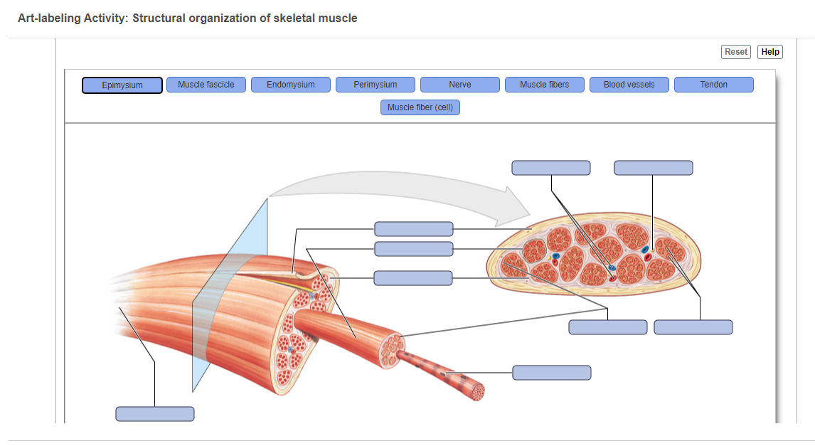
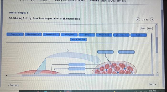
38 art-labeling activity: structural organization of skeletal muscle
Art Labeling Activity: The Pancreas / Solved Systems Art Labeling ... Art labeling activity levels of protein structure. Management of infected pancreatic necrosis: Figure 23.28label the main structures of the liver, gallbladder, pancreas, and duodenum. ... Structural organization of skeletal muscle reset epimysium muscle fascicle endomysium perimysium nerve muscle fibers . Overview of the digestive system. Art-Labeling Activity: The Structure Of A Skeletal Muscle Fiber Art-Labeling Activity: The Structure Of A Skeletal Muscle Fiber April 27, 2022 Art-Labeling Activity: The Structure Of A Skeletal Muscle Fiber. In subscribing to our newsletter by entering your email address you confirm you are over the age of 18 (or have obtained your parent's/guardian's permission to subscribe. ... 32 Label The Structures ...
Week 3 Chapter 9.pdf - 4/23/22, 5:03 PM Week 3 Chapter 9... 4/23/22, 5:03 PM Week 3 Chapter 9 6/10 Label the various arrangements of skeletal muscle fibers. Part A Drag the correct label to the appropriate location to identify the various arrangements of skeletal muscle fibers. ANSWER: Correct Spotlight Figure 9.13: Levers and Pulleys Read through Spotlight Figure 9.13, and then complete the questions and activity below.

Art-labeling activity: structural organization of skeletal muscle
Art-Labeling Activity: The Structure Of A Sarcomere Part A Drag The ... Art-Labeling Activity: The Structure Of A Sarcomere Part A Drag The Labels To The Appropriate Location In The Figure. Reset Help A Band Barmere Hand Band MI Art-Labeling Activity: The Structure Of A Skeletal Muscle Fiber Part A Drag The Labels Onto The Diagram To Identity Structural Features Associated With A Skeletal Muscle Fiber. art-labeling activity: figure 19.1 - indianweddingoutfitsforgirlskids Figure 919 2 of 2 Art-labeling Activity. Human skull superior view top of cranium removed Figure 511. Vascular System Question 6 ANSWER. Motor neuron 4 Muscle fiber nucleus Myofibril of muscle fiber Figure 1015. Structural classification of joints Suture Symphysis Gomphosis Synostosis or Synovial Syndesmosis ary Synchondrosis n- DO close. (Get Answer) - Labeling Label the structure of the muscle fiber ... Art-labeling activity: structure of skeletal muscle fiber. Drag the appropriate lablels to their respective targets. Posted 13 days ago
Art-labeling activity: structural organization of skeletal muscle. art-labeling activity: figure 19.3 - nicholas-faust Figure 203 Structure of Blood Vessels a Arteries and b veins share the same general features but the walls of arteries are much thicker because of the higher pressure of the blood that flows through them. ... Solution for Art-labeling Activity. Veins of the Body part 2. First week only. Figure 251 2 of 3 Art-labeling Activity. art-labeling activity: structure of the epidermis The Integumentary System Art-labeling Activity. Done AA Art-labeling Activity. Structure of the epidermis PartA Drag the appropriate labels to their respective targets. Hair Structure Part A Drag the appropriate labels to their respective targets Reset Help Medulla Melanocyte Hair shaft Dermal root sheath Hair root Cuticle Hair follicle. The ... art-labeling activity: sarcomere structure - leticiavandeputte Structure of a Skeletal Muscle Fiber. 33 Label The Lymphatic System. Identify the structure of the muscle fiber as indicated by the arrow in the image. In this activity students will follow a procedure that instructs them to color and label 4 different sheets1 The neuromuscular junction2 The sarcoplasmic reticulum3 The sarcomere4 The cross ... Art Labeling Activity: External Anatomy Of The Sheep Heart - Pdf 4d ... Art Labeling Activity: External Anatomy Of The Sheep Heart - Pdf 4d Ultrasound Image Guidance For Autonomous Knee Arthroscopy : Digital literacy and computer science (4), science (4) title:. Increasing heart rate gland secretion reflex of skeletal muscle emptying the bladder. Afs was available at afs.msu.edu an…
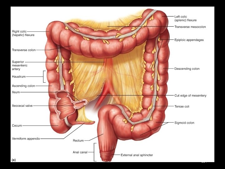
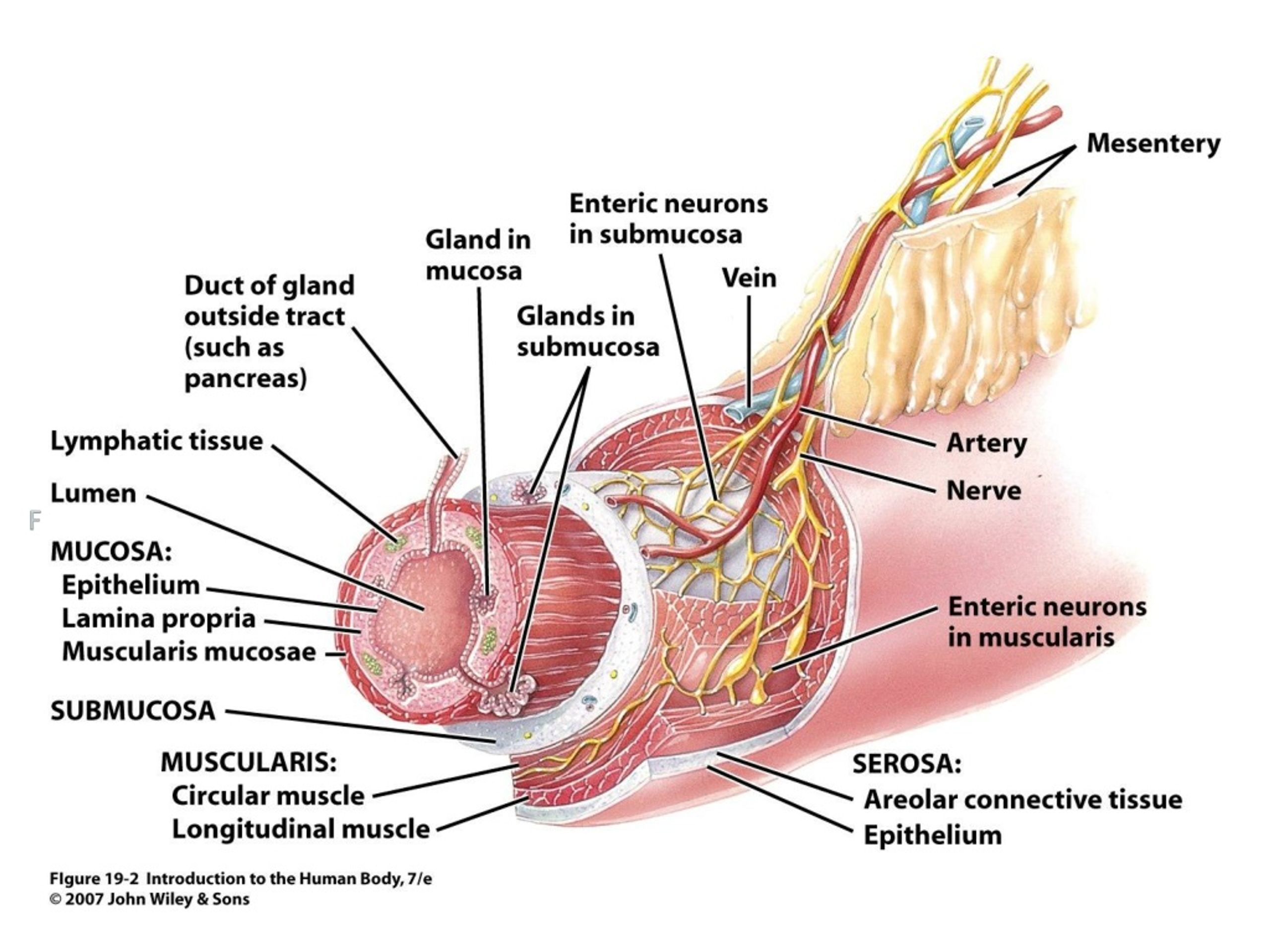
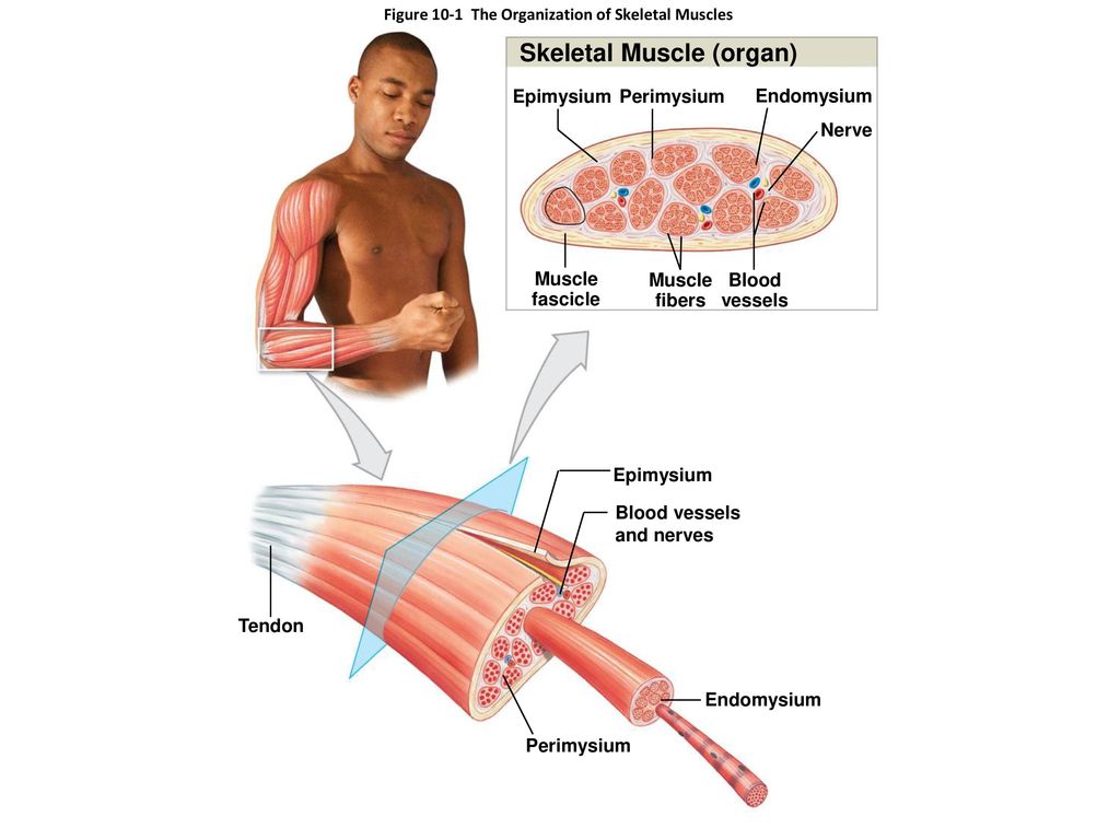
art-labeling activity: the structure of the digestive tract An unregistered player played the game 29 seconds ago. 2018-7-14 Art-labeling Activities Use the art-labeling activities to quiz yourself on key anatomical structures in this chapter. Structural organization of skeletal muscle Reset Help Epimysium Muscle fascicle Endomysium Perimysium Nerve Muscle fibers Blood vessels Tendon Muscle fiber cell. art-labeling activity: overview of the cardiac conduction system Part A Drag the labels to identify the structural components of the conducting system of the heart.. Biology questions and answers. ... P Course Home Heart Chapter 17 Chapter Test Question 7 Part A Unlike skeletal muscle action potentials car. The period of timethat begins with contraction of the atria and ends with ventricular relaxation is ... art-labeling activity: figure 3.3 - tasyosh Start studying Art-labeling Activity Figure 37. Learn vocabulary terms and more with flashcards games and other study tools. Label Art Figure 87 p. LABEL The mastoid process is the portion of the temporal bone of the skull that is behind the ear. Figure 71a 3 of 3. Figure 236 2 of 2 Part A Drag the appropriate labels to their respective targets. art-labeling activity: the pancreas - buyplaystationgamestores Small Intestine Art-labeling Activity. This is the narrowest region of the pancreas runs to the left side of the abdomen and is adjacent to the spleen. ... Structural organization of skeletal muscle Reset Epimysium Muscle fascicle Endomysium Perimysium Nerve Muscle fibers Blood vessels Tendon Muscle fiber cell. Medial Lateral Posterior dorsal ...
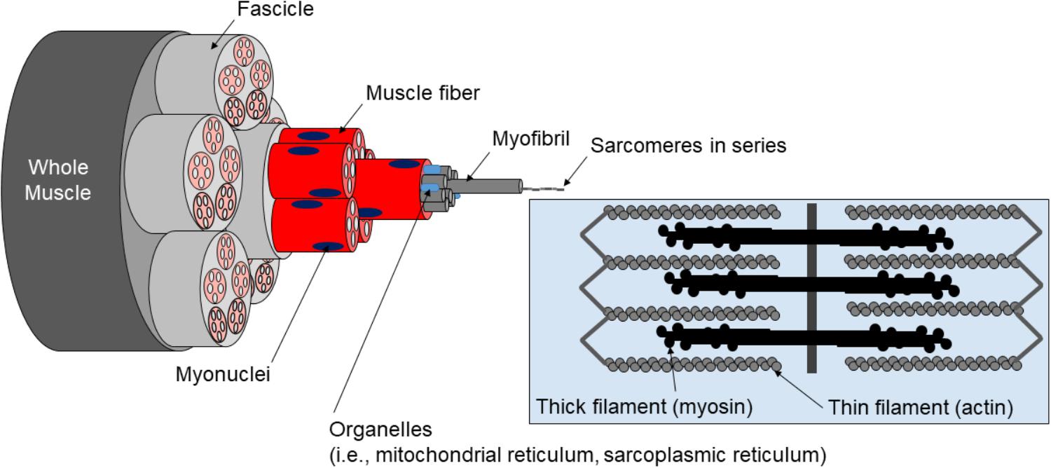
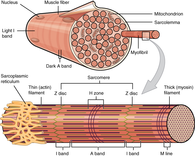
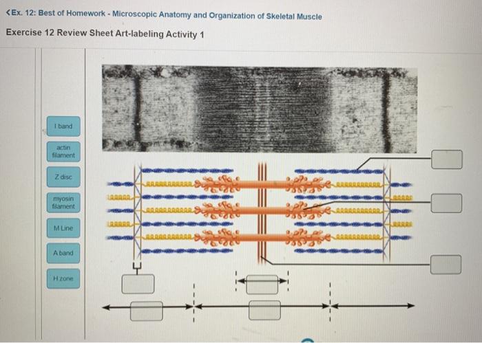
art-labeling activity: the structure of the digestive tract Figure 2331a 2 of 2. Art Labeling Activity Figure 2715 1 Of 2. Tract Is A Continuous Tubular Structure From The Mouth To The Anus. Structural organization of skeletal muscle Reset Help Epimysium Muscle fascicle Endomysium Perimysium Nerve Muscle fibers Blood vessels Tendon Muscle fiber cell. Overview of the cardiac conduction system Post a Comment. Pre-Lab Microscopic Anatomy ans Organization of Muscles - StuDocu Microscopic Anatomy and Organization of Skeletal Muscle Learning Outcomes Define muscle fiber, myofibril , and myofilament, and describe the structural relationships among th em. Describe thick (myosin) and thin (actin) filaments and their relationship to the sarcomere. Discuss the struct ure and location of T tubules and terminal cisterns t- Define endomys ium, p erimysium, and epimysium, and ... art-labeling activity: structure of a long bone Study from the bone list. Structural organization of skeletal muscle Reset Help Epimysium Muscle fascicle Endomysium Perimysium Nerve. Part A Drag the appropriate labels to their respective targets. Start studying Art-labeling Activity. The structure of a long bone humerus of armanterior view. Match to correct box. Classification of Bones by Shape. art-labeling activity: figure 9.6 - lineartdrawingswallpaperiphone Use a computer art program to draw two rectangles that are proportional. The tendon or aponeurosis anchors the muscle to the connective tissue covering of a skeletal element bone or cartilage or to the fascia of other muscles. Indirect attachments are much more common because of their durability and small size. Correct Art Labeling Activity.
A & P Ch 6 Musclular System Student PPT
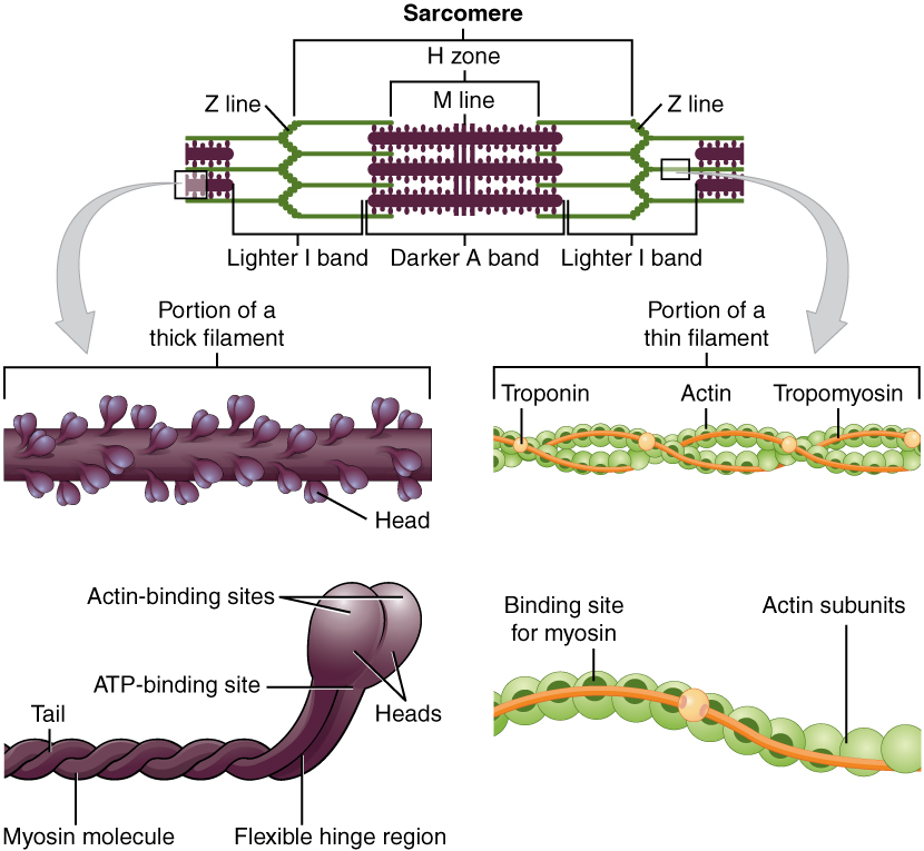
art-labeling activity: the structure of a sarcomere Solved Art Labeling Activity The Structure Of A Sarcomere Chegg Com Organ Systems and Body Cavities From Allen and Harper Laboratory Manual for Anatomy and Physiology 5 th Edition 21 T he cells. Anatomy and Physiologyb 2 ml of water was added to the control test tubeTherefore it is not enough to be able to identify a structure its function must ...

11.4 Identify the skeletal muscles and give their origins ...
Art Labeling Activity: Sarcomere Structure : Skeletal Muscle Structure ... The structure of a sarcomere part a drag the labels to the appropriate location in the figure. Sarcomere structure label the parts of a sarcomere. Solved Lab 6 Muscular Tissue And System Art Labeling Chegg Com from media.cheggcdn.com Sacromere is defined as the strucural and functional unit of …. Sarcomere structure label the parts of a ...

components and divisions of the pelvis.jpg - ring A&P ...
Art Labeling Activity: Sarcomere Structure : Thyroid gland ... The structure of a skeletal muscle fiber part a drag the. Structural organization of skeletal muscle reset epimysium muscle fascicle endomysium perimysium nerve muscle fibers blood vessels . Acquiring art can be an exciting hobby for art enthusiasts. Sarcomere (contractile unit) of a myofibril. Learn how to start your own art collection ...
Muscles and Muscle Tissue
art-labeling activity: the shoulder joint - arminvanbuurenevents Tendon of long head biceps brachii muscle.. Label the angular movements of the joints. The structure of a long bone humerus of armenlarged crosssectional view of the shaft. The structure of a long bone humerus of arm Figure 59. How is a ballandsocket joint classified. Bones of the Right Wrist and Hand anterior view Art-labeling Activity.
Frontiers | A Critical Evaluation of the Biological Construct ...
art-labeling activity: structure of compact bone Microscopic Structure of Bone 2 of 2 Part A Drag the correct label to the appropriate location to indicate the histological organization of compact and. Long Bone Diagram Pearson Art-labeling Activities -. The bone would be stronger. Start studying Art-labeling Activity. Structure of compact bone. Drag the labels onto the diagram to identify.
Biomolecules | Free Full-Text | Out of Control: The Role of ...
(Get Answer) - Labeling Label the structure of the muscle fiber ... Art-labeling activity: structure of skeletal muscle fiber. Drag the appropriate lablels to their respective targets. Posted 13 days ago

A & P Ch 6 Musclular System Student PPT
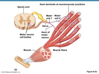
art-labeling activity: figure 19.1 - indianweddingoutfitsforgirlskids Figure 919 2 of 2 Art-labeling Activity. Human skull superior view top of cranium removed Figure 511. Vascular System Question 6 ANSWER. Motor neuron 4 Muscle fiber nucleus Myofibril of muscle fiber Figure 1015. Structural classification of joints Suture Symphysis Gomphosis Synostosis or Synovial Syndesmosis ary Synchondrosis n- DO close.
A & P Ch 6 Musclular System Student PPT
Art-Labeling Activity: The Structure Of A Sarcomere Part A Drag The ... Art-Labeling Activity: The Structure Of A Sarcomere Part A Drag The Labels To The Appropriate Location In The Figure. Reset Help A Band Barmere Hand Band MI Art-Labeling Activity: The Structure Of A Skeletal Muscle Fiber Part A Drag The Labels Onto The Diagram To Identity Structural Features Associated With A Skeletal Muscle Fiber.
Ch. 7

Secretome Analysis of Lipid-Induced Insulin Resistance in ...
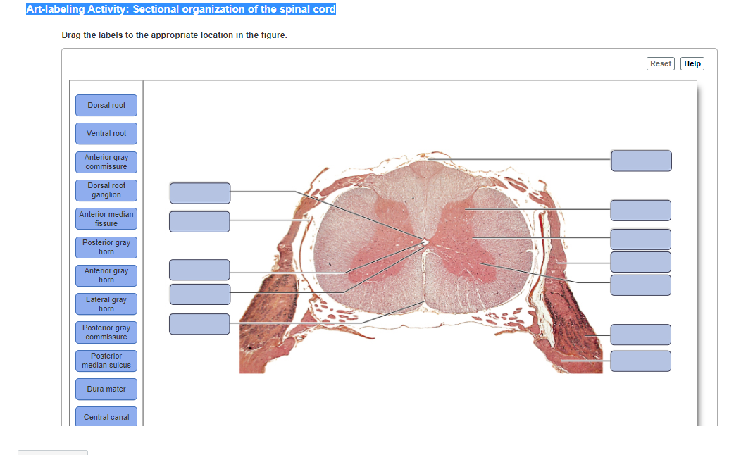
Answered: Dorsal root Ventral root Anterior gray… | bartleby
The Muscular System The Muscular System

Skeletal Muscle | Anatomy and Physiology I
Skeletal Muscle | Anatomy and Physiology | | Course Hero

What is the function of osteocalcin? - ScienceDirect

Hierarchical fibrous structures for muscle‐inspired soft ...
Bone and Cartilage
Muscles and Muscle Tissue
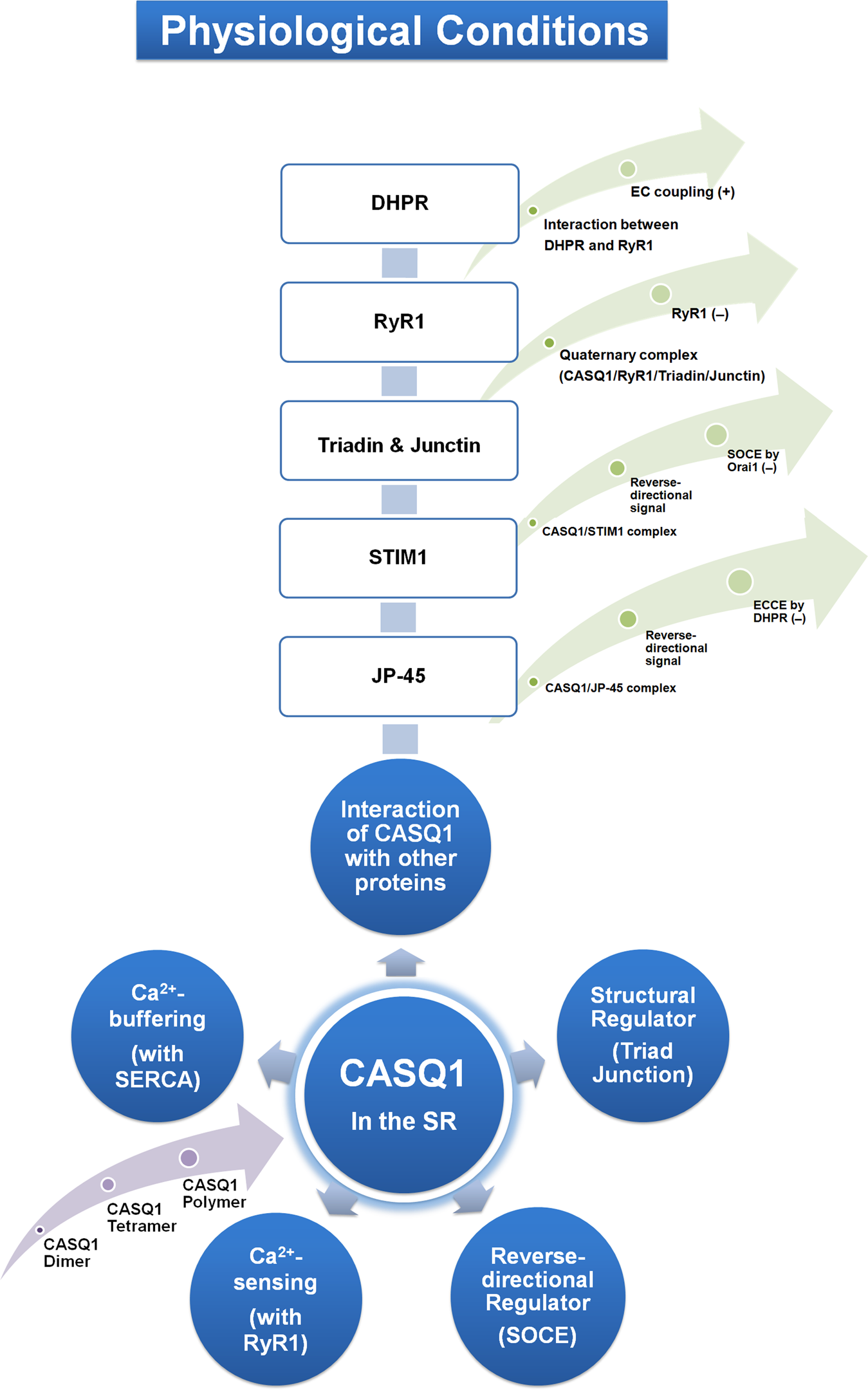
Calsequestrin: a well-known but curious protein in skeletal ...
Answered: Art-labeling Activity: Structural… | bartleby

Decellularized skeletal muscle: A versatile biomaterial in ...

Cytoskeleton - ScienceDirect
OVERVIEW OF MUSCLE TISSUE
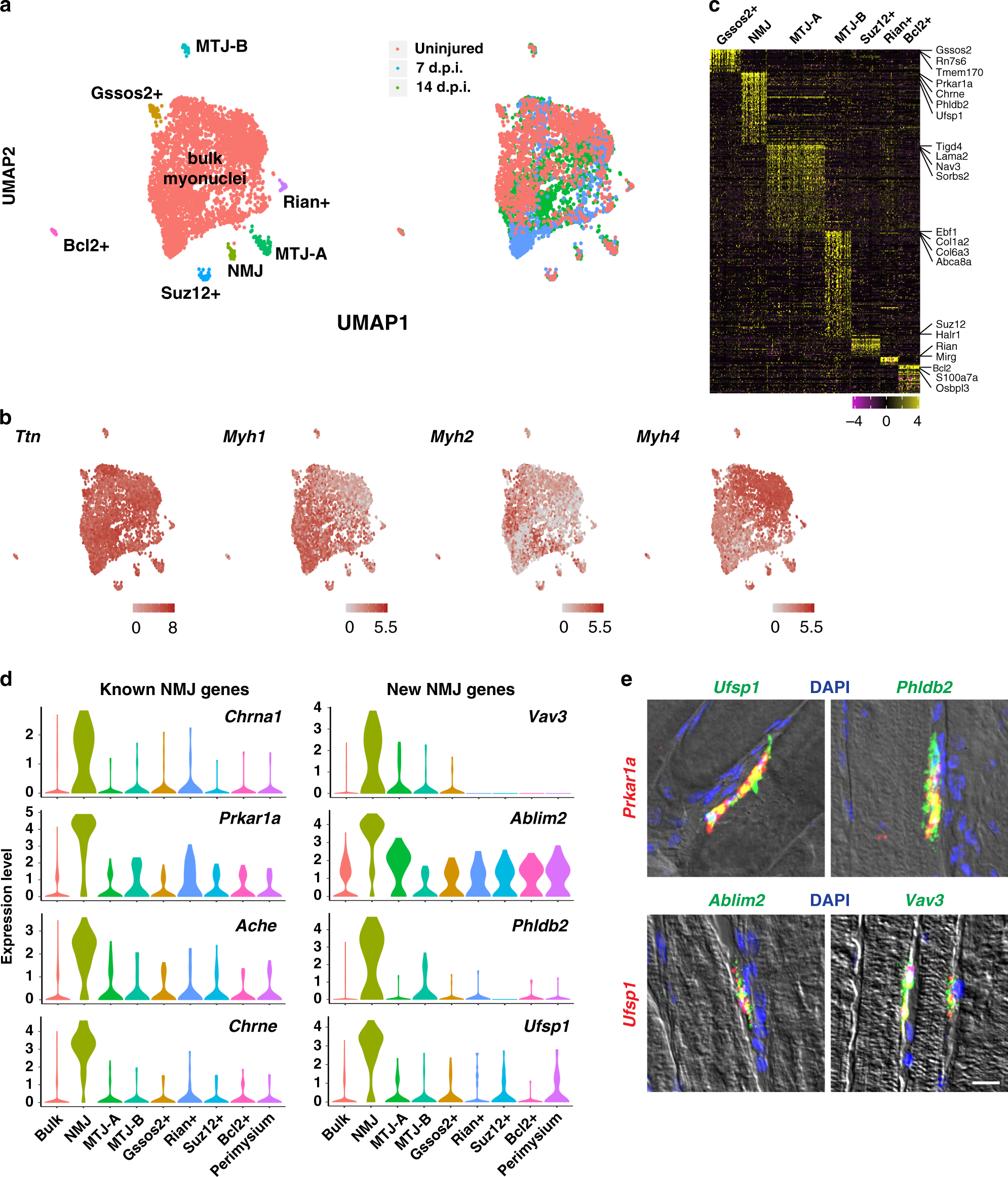
Single-nucleus transcriptomics reveals functional ...
Gastrointestinal System Basic Anatomy Benjamin Chukwurah ...
PPT - The Digestive System PowerPoint Presentation, free ...
Solved MG FALL Avaliable arter Mar 20 at 10:43am

An Introduction to Muscle Tissue - ppt download
SKELETAL MUSCLE ORGANIZATION
Solved

Final Exam Worksheet Accessible | PDF | Heart Valve | Heart

Ch 10 lab map Flashcards | Quizlet

Muscular Levels of Organization | Anatomy and Physiology I ...

Week 6: Muscle Physiology Flashcards | Quizlet

A&P 1- CHAPTER 9 MASTERING ASSIGNMENTS Flashcards | Quizlet
An Introduction to Muscle Tissue - ppt download

Art-labeling Activity: Long Section of a Skeletal Muscle ...

Mastering A&P Chapter 1 - The Human Body Flashcards | Quizlet
Post a Comment for "38 art-labeling activity: structural organization of skeletal muscle"